florida joe
Well-Known Member
from the web
Fragmentation of an Elegance Coral, Catalaphyllia jardinei
Fragmentation is common in stony corals that contain large numbers of colonial polyps (Acropora,Montipora, etc.) and in those with multiple individual polyps such as phaceloid forms of Euphyllia. On the other hand, stony corals that exist as fused or conjoined polyps such as Catalaphyllia,Plerogyra, Fungiids, etc. present several special challenges, and are generally not regarded as good candidates for fragmentation. Many solitary polyped corals generally have heavy, bulky skeletons which are more challenging to cut. There is also the very understandable concern of losing the entire animal if the fragmentation is not tolerated. Of course, this is the case with any propagation effort. But, it would seem that the risk is perceived to be greater among aquarists when dealing with these types of corals. It appears that the comfort level is high when it comes to breaking or cutting the ‘little sticks’ that we grow, as evidenced by the multitude that are available for trade or sale, but talk to a group of hobbyists about cutting through a large fleshy polyp and they usually become very squeamish. Additionally, fragments of large polyped corals are thought to take longer to grow into “normal looking” marketable colonies, but that is not the case with Elegance corals as one will quickly see.
Several factors motivated the fragmentation of this particular specimen. The coral had grown too large for the space available to it in the aquarium and had begun to cause significant damage to neighboring corals with its stinging nematocysts. Also, because of the recent apparently poor survival rates of freshly imported Catalaphyllia, it is our hope that this demonstration will encourage thoughtful propagation of healthy specimens that have been successfully kept in captivity for some time, while sparing wild collected animals an almost certain death. Last, but certainly not least, healthy specimens of Catalaphyllia were needed by Eric Borneman for his Elegance Coral Project,) which seeks to identify the causes of this coral’s currently poor survival rate.
Although the fragmentation procedure that was chosen for this coral is fairly simple, a lot of planning went into the process to maximize both pieces’ chances of survival. The following paragraphs outline the steps taken and their rationale.
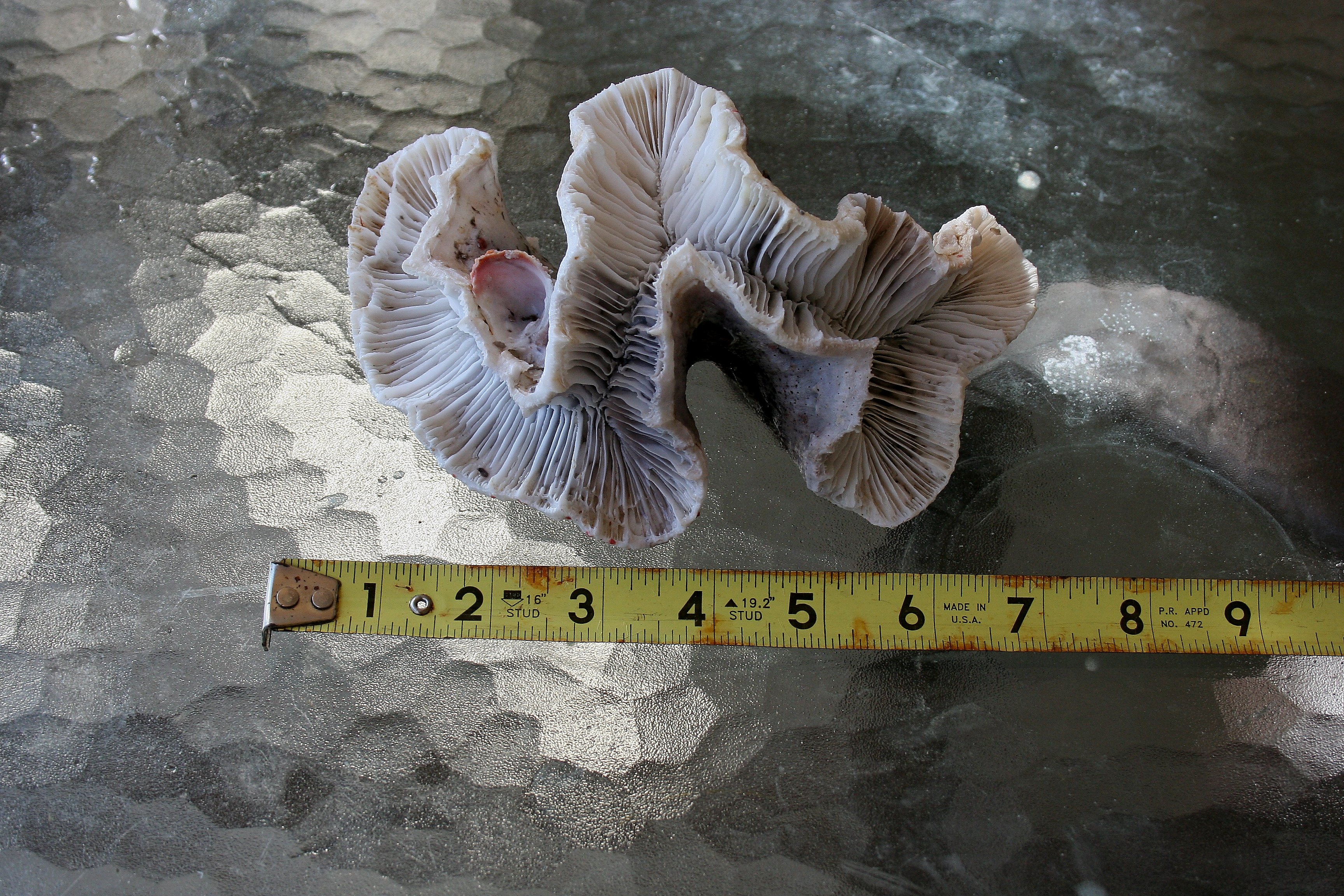
Before removing the coral from the aquarium, it was gently “waved down” and shaken in order to retract its polyps, to prevent damage to the polyps caused by the weight of its own water-filled tissue when removed from its aqueous environment. Note the ridge on the skeleton where the growth pattern changed. All growth above this ridge occurred during the 16 months when the coral was in the care of Adam Cesnales.
The coral was placed in a flat tub that was deep enough to cover it, but shallow enough to work easily. This allowed the coral to be manipulated and cut with its skeleton out of the water, but with its tissue remaining submerged.
Both authors recalled reading reports on the Internet of an interesting technique for propagating wall-type large polyped stony corals such as this, but it would appear that the original webpage has disappeared in cyberspace. In these reports, the skeleton was divided, but not the living tissue. A wedge was then placed between the parts, and the connecting tissue was allowed to separate on its own. This technique was obviously born out of fear of cutting large amounts of living tissue. We decided that this would serve only to impede water flow to the tissue that inevitably would be damaged in the process, so this plan was rejected. Causing as little tissue damage as possible remained an important goal, so a significant amount of time was invested to choose the best place to cut. A part of the coral was chosen that would be both accessible to the cutting tool and that would render two attractive coral pieces, while at the same time minimizing the disruption of living tissue.
When a location was chosen for the cut, the same type of rotary tool equipped with a side-cutting bit was first plunged through the skeleton near the polyp. The cut was then extended outward, away from the tissue toward the edge of the skeleton. This offered much greater control than trying to initiate the cut from the edge of the skeleton and then moving toward the polyp. Also, this type of cutter (as opposed to a disc) allowed the skeleton to be cut all the way through in a single pass, thereby making a cleaner cut. The skeleton was much more friable than expected and the cutting tool passed through it with minimal resistance.
The cut was then extended toward the polyp. A bulb syringe was used to irrigate the cut, clearing away the grit and making it easier to see. The intent was to cut most of the way through the skeleton and then break the last 1-2cm, thus avoiding contact between living tissue and the cutting tool. While still cutting about 3-4cm from the polyp, however, a white pasty substance began to run from the cut. Since living tissue obviously had been encountered more deeply into the skeleton than expected, the cutter was stopped and the remaining skeleton was broken.
Once the skeleton was separated, a scalpel was used to carefully cut the tissue connecting the two pieces. Mesenterial filaments, as well as other tissue, were clearly visible at the margins of the cut, and the living tissue extended surprisingly deeply into the skeleton. As the reader can imagine, a prized coral with its “guts” hanging out is not a comforting sight!
The new fragments were returned to the original display and placed as closely to their original location as possible to avoid any additional stress from changes in lighting or current. The fouled water that contained the coral during the fragmentation process was discarded. The authors then took a short lunch break and in the amount of time it took us to consume some pizza, chicken wings, and a few beers, both portions of the coral had expanded to nearly normal size. This image was taken about five hours after fragmenting the coral. Cleary, the procedure was tolerated well! Within about two weeks, new tissue had covered the cut edges.


